Tethered to science


Tethered to science

For a long while I haven’t posted anything, being too tired and work got in the way. Then after combing back from a conference and a break late February, full of ideas but no energy to work them out, I turned to my GP.  There I was told I was overworked, warned that if I did not slow down I will end up with a burn out. So I tried to rest and slow down, while still having that nudge of guild when leaving work early. Then the corona crisis hit. Forced to work at home made me feel less guilty for the times I stopped earlier, because hey we where in a crisis, so its ok not to be able to concentrate. Needless to say it did wonders for my recovery.

Working on a review article that was already in the pipeline, I recovered slowly. Not only am I again able to work a full day without loosing my concentration. I also found back what I lost long ago. How much I enjoyed just reading articles, being able to follow up on what I read, while discovering how it all worked. Then finding a way to describe this so others would see the connections between the different studies as well.

In short it showed me that biology is amazing and has find some inventive solutions to its problems.

One of these are tethering proteins. These are proteins that are connected to membranes of different organelles, say the endoplasmic reticulum (ER) and the plasma membrane, to keep them close to each other. Tethering proteins make use of two different domains. On one end of the protein they have a transmembrane domain. This domain insert itself through the membrane to anchor the protein to it. On the other end they have a couple of domains that can bind membranes.

Tethering protein
A schematic overview of a tethering protein, TM: transmembrane domain, C2: C2 domain.

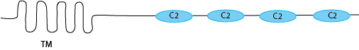
There are lots of different types of domains that can do that. But there is one in particular that is quite interesting in this context, the C2 domain. The C2 domain binds to membranes in a calcium dependent manner. But the concentration of calcium required depends on the exact sequence, so there are C2 domains that need just a little bit of calcium and they will already bind membranes. Then there are others that need quite a high calcium concentration for them even have a chance to bind the membrane. Tethering proteins make use of these C2 domain characteristics. They have a low calcium requirement C2 domain at the extreme end of the protein, and then another, one, two or three C2 domains, each requiring a bit more calcium before they are able to bind the membrane, placed more towards the transmembrane domain.

tethered membrane low Ca2+
A tethering protein holding two membranes together under low calcium conditions

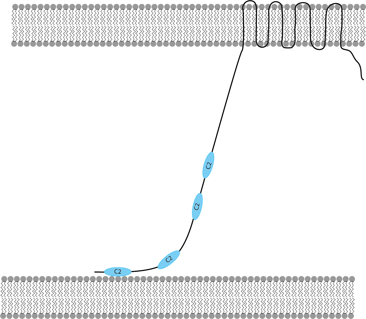
In this way, by using tethering proteins, the cell can hold both, say the ER and the plasma membrane, just having a little bit of calcium present.

Tethered membrane high Ca2+
A tethering protein holding two membranes together under high calcium conditions

But when the ER and the plasma membrane needs to be really close, say for some signalling function, then the cell can increase the calcium concentration, and the tethering protein will just reel in the ER close to the plasma membrane.

That image of the ER tethered via a leach to the plasma membrane, so it can be brought in close when needed. Is just one of the many I got over the past weeks, reading for my review. I will share some more over the coming weeks, illustrating how science reeled me in once again.

Literature

Brault ML, Petit JD, Immel F, Nicolas WJ, Glavier M, Brocard L, Gaston A, Fouche M, Hawkins TJ, Crowet J-M, et al (2019) Multiple C2 domains and transmembrane region proteins (MCTPs) tether membranes at plasmodesmata. EMBO Reports 20: e47182

Ishikawa, K., Tamura, K., Fukao, Y. and Shimada, T. (2020), Structural and functional relationships between plasmodesmata and plant endoplasmic reticulum–plasma membrane contact sites consisting of three synaptotagmins. New Phytol, 226: 798-808.

Jiménez JL, Smith GR, Contreras-Moreira B, et al. Functional recycling of C2 domains throughout evolution: a comparative study of synaptotagmin, protein kinase C and phospholipase C by sequence, structural and modelling approaches. Journal of Molecular Biology, 333(3):621-639.

Plasticity in phloem development


Plasticity in phloem development

Last week at a symposium, we were reminded by Antia Rodriguez-Villalon that in plants organogenesis does not stop after germination. In fact, plants keep producing new organs through their lives. While most of us think by organ formation in plants first about leaves or flowers, Antia Rodriguez-Villalon work actually focusses on vascular development in roots. Her main take home message was that vascular development is more plastic than we initial thought. And that this plasticity safeguards the development of a functional vascular system. So I was excited when this weekend I saw in a tweet about the latest article of her group describing this study.

protophloem - root
The radial organization of the vascular tissues in Arabidopsis roots where depicted phloem tissues are color coded (CC, companion cell; MSE, metaphloem sieve element; PPP, protophloem pole pericycle cells; PSE, protophloem sieve element). Copied from Gujas et al. 2019

Before I go into more details about her work we will take a short detour, about phloem development. In plants the fate of a cell is determined by its position. As such, we know the function of a cell by its position in the plant. In Arabidopsis roots the phloem pattern is made up of four cell types, and is well conserved. With the protophloem and the metaphloem sieve elements originating from a common stem cell, whereas the companion cell originates from a different stem cell. In effect, once the precursor cells to the protophloem and the companion cells get pushed outside the meristem region, these cells are in different but adjacent cell files. Cells that have a protophloem identity can be visualised with a protophloem marker.

Using this technique they looked at how the protophloem identity was affected in the mutant, cvp2 cvl1, which is severely compromised in protophloem development. Finding that protophloem identity was affected, they set out to determine the new identity of these affected cells. Surprisingly, these affected cells had a gene expression similar to that of companion cells. Investigating this a little further, by intentionally disrupting the protophloem development, showed that in case of disruption the protophloem identity switched from the protophloem cell-file to the companion cell cell-file. Giving the first hint of the plasticity of the protophloem development. This plasticity is restricted to a so called “plastic zone” in which the cells surrounded the protophloem are still in an uncommitted stage. Eventually, through growth, these uncommitted cells will be pushed out of the plastic zone and commit.

Further study to investigate how the plastic zone is regulated identified that RPK2 and CLE45 function to restrict protophloem identity to the protophloem position. In addition, CLE45 treatments prevented the plasticity of protophloem development. Suggesting that in the plastic zone, plants normally can modulate CLE45 perception at single cell level, enabling them to re-pattern to form a functional phloem pole upon positional cues.

protophloem plastic zone schematics
Schematic Overview of the Molecular Mechanisms Regulating PSE Identity and Phloem Patterning in Arabidopsis Roots. A longitudinal view (left) and radial view (middle) of the developmental trajectories of protophloem sieve elements (PSEs) and companion cells (CCs) within the root are represented. Previous to their entry into the plastic zone (surrounded in red), future PSE cells acquire their cell identity and enter into a proliferative phase, a process partially regulated by the activity of positive regulators (such as CVP2) and counteracted by negative regulators, such as CLE peptides. Within the plastic zone, PSE-surrounding elements are primed as phloem cells, but they still exhibit plastic identity and can switch their identity (red arrows) according to positional cues. Once PSE cells enter into differentiation process, RPK2 excludes PSE identity from PSE-surrounding cells, allowing these cells to commit to CC’s developmental trajectory. Copied from Gujas et al. 2019

This latest work of the group of Antia Rodriguez-Villalon showed us that phloem development has a back up plan for it worse case scenario. I look forward for them to find out more about how this is organised.

Literature

Gujas et al., A Reservoir of Pluripotent Phloem Cells Safeguards the Linear Developmental Trajectory of Protophloem Sieve Elements, Current Biology (2019), https://doi.org/10.1016/j.cub.2019.12.043

What is wrong with how and when we are taught to write grants


What is wrong with how and when we are taught to write grants

Doing science for a living is one of the most fantastic jobs you can think about. And in general we scientist have a pretty good deal, being at the forefront of new discoveries and getting paid for learning new things. However, for all the great things that go along with it, job security is not one of them. Most of us have faced the situation of being in a great place, working on a  fantastic project, dreaming of making a home in this place, when the reality hits, our contracts run out and without any new grant on the way they have to let you go.

Is it then not strange, that for a sector so dependent on grant money coming in, that scientist are hardly thought how to write grants. As students, all up to the end of our PhDs and even when being postdocs we learn how to do science. From the most basic of how to make up solutions, maintain cell lines, and germinating seeds. To designing and executing complex experiments aimed at answering our questions. We get thought how to write down our results, how to report them to others. Both in writing and in presenting those results in meetings and conferences. During our PhD and while doing our postdocs we get opportunities to supervise others, learning how to lead. We get chances to review papers and to scrutinise the work of other scientists. But the one thing, the most crucial thing on which science relies, applying and with luck receiving a grant to do the research that we are dreaming of, is not thought in any reliable way.

Yes if you are lucky, you might have had an assignment of writing a grant when you were doing your bachelors or master degree. But, who will remember that when they are in the middle of a second postdoc trying to figure out how to get into that magical kingdom in which PIs seem to live.  When you are part of a well financed lab, you would have no need to apply for any kind of grant up to the point that you decide that you have that great idea and want to start your own lab. To be honest, this is not great timing, being thought how to write and apply for a grant or a fellowship at the moment your career depends most on it. It is the one and foremost thing postdocs ask for, teach us how to write and apply for grants. Grant writing courses for postdocs are filled up in no time. Because we know our careers depend on them.

I would suggest a change of practice. Not only should PIs encourage PhDs and Postdocs to apply for travel grants. They also should start a policy in which all new postdocs, will need in their first year, write and apply for a grant or fellowship. In this way postdocs get the training in writing and applying for grants without any real losses when they don’t get them, but with big wins when they do. For those who win the grant it might buy them an extra year or two on their contract. Enough time to cash in on their hard work in the form of some nice papers and preliminary data which they can use in their application for their next grant.

If at the same time PIs introduce a second standard policy, by asking postdocs, for example during those annual review meetings, what they want to do next. And then, of course, not file this information away, but actively help to work out multiple ways for the postdoc to get there, irrespective if this in academia or outside. It might be a way for postdocs to land that fabled staff scientist position they have heard about but never seen advertised. It might be how a postdoc find themselves on an institutes magazine on their way to become and actual science journalist. It might be how as a postdoc you take your results outside the lab and into your start up. And, who knows, it might even be the way how a postdoc finds it way into the magical kingdom of the PIs.

I know this sounds like a lot of extra work for PIs. But, once the first year of postdocs have been initiated in the art of grant writing they are able to help the next who arrives. The same with knowing the passion of your lab members, once known they can be applied when needed. Benefitting both the lab and the person doing it.

Leaf shape development


Leaf shape development

On of the things that intrigues me most in biology is the development of organisms. How does that single cell that is just fertilised knows what to do. To get its polarity established, initiate cell division at the right time, place and direction. What makes it go on developing into recognisable plant and not just a mass of cells. It simply fascinates me. I guess that is what is exciting of the research coming from Enrico Coen lab. I was first introduced to what his lab was doing while working at the John Innes Centre, through our departmental seminars and the annual research days. Here I was introduced to the concepts of how you could compute the development of a leaf.

This they did through painstakingly following the developments of Arabidopsis leaves, tracking cell divisions, from the tiny leaf primordia up to the fully grown leaf. Studying the relationship between cell division, cell size and growth rate. This information they then used to feed into computational models. Which as explained in a recent publication that leaf shape could be brought back to a few parameters and growth factors. The important parameters included growth rate (perpendicular and proximodistal) division rate (division competence and mean threshold cells size). And the growth factors could be brought back to a graded proximodistal factor (PGRAD), a mediolateral factor (MID), a factor distinguishing lamina from petiole (LAM), timing factor (LATE),  and proximal mobile factor (PMF). Interestingly by slightly tweaking the influence these growth factors have an effect on the leaf growth the size of the leaf. This is corresponding to what is observed in mutants with a leaf size phenotype. Another exciting part of this research is that it shows that it is possible to obtain variation in leaf size with just a defined set of growth factors, corresponding to transcription factors in the plant.

These initial studies focused mainly on simple leaves, flat and round. But now they taken this to a whole new level, going from a 2D to a 3D leaf shape. There they show how carnivorous plant carnivorous plant Utricularia gibba adjust their planar (simple) leaves into needle-like structures and cup-shaped traps. First using the same through approach  they used 3D imaging to track cell divisions and to obtain growth rate measurements in three dimensions to use to build a model of how the cup-shaped traps develop. This resulted in a model similar to that of an Arabidopsis leaf, with parameters for growth rate and division rate and growth factors representing a mediolateral factor (MID), ventral midline factor (VEN), and a stalk diameter factor (STK). Using the observed parameters they showed that model was able to reproduce the observed development of an Utricularia gibba cup-shape trap. In addition they showed that by adjusting the parameters they could generate trap morphologies similar to other Utricularia species.

However, a limiting point of the model is in that it considers only the later stages of development of the trap. Not explaining how the curvature of the trap primordium originates. One way of how the initial curvature for the cup-shaped trap might be initiated from a two dimensional origin is through abaxial and adaxial patterning. Comparing the cup-shaped traps with simple leaves it was deducted that the outer side of the cup corresponds to the abaxial (lower) side, and the inner side to the adaxial (upper) side of a simple leaf. Therefor the latest work of the Coen lab investigates the influence of the effect of adaxial and abaxial domains on the polarity field that orientates growth. The decision if cells are part of the adaxial or abaxial site of the leaf occur via gene activity in the leaf primordia. However, solely based on leaf morphology you would not able to distinguish between future leaves or future traps as they are all dome-shaped.

To understand how adaxial and abaxial domain specification might influence trap development the adaxial-abaxial domains in developing traps were identified. While in primordia of leaves the adaxial and abaxial domains each take up approximately halve of the dome-shaped primordia, in primordia of traps the adaxial domain was much smaller than the abaxial domain. To investigate the influence of this on trap development, they build a model in which they could specify the adaxial and abaxial regions. Finding that having an equal adaxial-abaxial distribution would result in either simple leaves or needle like leaves, depending of the settings of the parameters for the different growth rates. However, when they restricted the adaxial region to only a small region on one side of the primordia they found that the resulting leaf would develop into a cup-shaped trap. Furthermore, by adjusting the size and form of the adaxial region, traps of different shapes could be generated.

With developing these models the Coen lab has illustrated the universal mechanism that is behind leaf development. The possibility to generate completely different leaves by adjusting only a few parameters points towards a small group of transcription factors whose differences in expression and activity between species might explain the variation in leaf shapes we see.

Finally, even if you did not understand much about what I have been talking about in this post, I would like to point out that they made lots of movies, both of the models as well as of the imaging they did in order to obtain all the data to feed into the models. You can have a look in the supplemental data of the discussed papers, as well as on the Coen lab website.